High risk pregnancy – Pregnancy is one of the most exciting and happy phase of women’s life. But for some, especially those with chronic medical conditions or who are expecting multiples, pregnancy management can be a time of intense fear and uncertainty. It is in those circumstances that we need to provide specialized care for both mother and child to ensure good health for both of them.
It is only in less than ten percent of pregnancies, which will have some kind of complication that can affect the health of the mother or the child. These are called as high risk pregnancy. When babies are born preterm, they have a higher risk of having serious health problems. If these high risk pregnancy can be identified and the pregnancy managed accordingly, they will be able to achieve a favorable outcome and they can have a comfortable pregnancy. In fact, many risk factors can be identified even before conception occurs
For all this they will require a comprehensive approach to their medical condition and pregnancy care along with a strong support system.
Common Risk Factors – High Risk Pregnancy
- Advanced Maternal Age
- Chronic medical conditions, such as lupus, cancer, diabetes , high blood pressure or arthritis
- Systemic complications in pregnancy
- Heart disease
- Renal disease
- Respiratory disorders
- Hepatobiliary dysfunction
- Haematological problems
- Family history of mental retardation or birth defects
- Women who have experienced miscarriages, pre-term deliveries, stillbirths, or neonatal deaths
Our Aim
The department of obstetrics and gynecology along with the feto maternal unit provides comprehensive care for high risk expectant mothers and their unborn babies. We provide a hospital based outpatient program that follows a multidisciplinary team approach.
At CIMAR , our team of experienced healthcare professionals is dedicated to offering comprehensive, compassionate care that includes
- Diagnosing potential pregnancy complications and fetal development issues in their early stages
- Providing top-of-the-line medical treatments to both mothers and infants
Being compassionate and offering emotional support during trying times
Facilities Available
The staffs are committed to providing comprehensive, integrated care for diagnosing, treating and managing high risk pregnancy all within a single, family oriented centre.
At CIMAR , our team of experienced healthcare professionals is dedicated to offering comprehensive, compassionate care that includes
The services include but are not limited to- Preconception and genetic counselling
- Prenatal testing and diagnosis
- High risk pregnancy consultation and maternity services
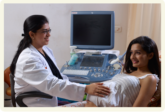
- Comprehensive 2-D, 4-D & Doppler ultrasound
- Diabetes education and nutritional counselling
- Lactation support and consultation
- Psychosocial assessment and lifestyle management
- Patient and family education classes and support groups
A high risk pregnancy diagnosis shouldn’t automatically have a negative connotation. With proper care, 90 to 95 percent of high risk pregnancy produce healthy, viable babies. The earlier a problem is detected, the better the chances that both mother and baby will stay healthy. It is important to remember, however, that not all conditions can be diagnosed, and some pregnancies begin normally, but develop problems later. Make sure you schedule regular visits with your doctor, before and after becoming pregnant.
Fetal medicine specialist is the specialist within the field of obstetrics. They deal with pre-conception counselling, especially in anticipated high risk cases both maternal and fetal point of view, offer prenatal tests (invasive and non-invasive), provide treatments, and perform surgeries. They act both as a consult during lower-risk pregnancies, and as the primary obstetrician in, especially, high-risk pregnancies. The perinatologist may work closely with paediatricians or neonatologists after the birth. Primatologists assist with pre-existing health concerns, as well as the complications caused in the pregnancy of the expectant mother.
Fetal medicine specialist, in addition to a degree in obstetrics have training in obstetric ultrasound, doing invasive prenatal diagnostic techniques like amniocentesis, chorionic villus sampling, fetal blood sampling and the management of high-risk pregnancies. Some are further trained in the field of fetal diagnosis and prenatal therapy where they become competent in advanced procedures such as targeted fetal assessment using ultrasound and Doppler, fetal blood sampling and transfusion, fetoscopy and open surgery for fetal.
What is a Fetal medicine ?
Fetal medicine (MFM), also known as perinatology is a branch of science. It focuses on managing health concerns of the mother and fetus before, during and shortly after pregnancy and delivery.
They take care of pregnant women who have (e.g. heart or kidney disease, hypertension, diabetes, and thrombophilia), pregnant women who are at risk for pregnancy-related complications (e.g. preterm labor, pre-eclampsia, and twin or triplet pregnancies), and pregnant women with foetuses at risk. Foetuses may be at risk due to chromosomal or congenital abnormalities, maternal disease, infections, genetic diseases and growth restriction.
For mothers, pre-existing health conditions, such as high blood pressure, drug use during or before pregnancy, or a diagnosed medical condition, may require consultation with a Fetal medicine specialist. In addition, women who have recurrent pregnancy loss may be referred to a Fetal medicine specialist for assistance.
During pregnancy, a variety of complications of pregnancy can arise. Depending on the severity and the complication, a Fetal medicine specialist may meet with the patient frequently and may become the primary obstetrician for the pregnancy. Post-partum, Fetal medicine specialists may follow up with a patient and monitor any medical complications that may arise.
The Society for Fetal medicine (SMFM) is an association of specialists who works to improve the maternal and child outcomes by standards of prevention, diagnosis and treatment through research, education and training.
Need for preconception and prenatal care and what they are in relation to Fetal medicine?
Preconception care – you may have to see a Fetal medicine specialist at least 3 months before planning a pregnancy especially in case of high risk pregnancy conditions which includes maternal conditions like hypertension, diabetes, heart disease, thrombophilia, epilepsy, prolonged drug intake for any medical condition, auto immune disorders etc., previous bad obstetric history, previous anomalous baby, history of genetic abnormality in the family/parents to provide a holistic approach to provide a positive outlook to a couple planning pregnancy so as to enter the pregnancy in optimal health to reduce antenatal fetal and maternal complications, thereby adding the need and importance of preconception care.
Genetic screening, ultrasound prenatal care and diagnosis
Screening for chromosomal or genetic abnormalities during pregnancy as a part of prenatal care is done by ultrasound examination as well as a series of blood investigations. Screening for Down syndrome should be offered for all pregnant women irrespective of the maternal age.
Diagnostic amniocentesis and prenatal care
Amniocentesis detects chromosome abnormalities and genetic disorders. Down syndrome or Trisomy 21 is the most common chromosome abnormality. Genetic disorders include disorders such as cystic fibrosis. Hence a diagnostic amniocentesis is indicated when screening for Down syndrome is positive, either of the parents/ previous child with any genetic or chromosomal disorder, ultrasound features suggestive of fetal anomalies.
Amniocentesis is usually performed between 16 and 20 weeks but can be done till 24 weeks. Some medical facilities may perform amniocentesis as early as 14 weeks. Very rarely it is done even in the third trimester in certain rare cases.
The best time to visit a specialist for diagnostic amniocentesis is hence before 16 weeks so that the test is properly planned.
Therapeutic amniocentesis
Therapeutic amniocentesis was defined as an attempt to remove enough amniotic fluid (Amnioreduction) in pregnancies complicated by symptomatic hydramnios to leave a normal volume of amniotic fluid (AF index less than 25 cm). It is also done in conditions where a drug needs to be delivered to the fetus directly for example in case of fetal hypothyroidism.
Therapeutic amniocentesis can be done throughout pregnancy irrespective of the gestational age as and when indicated.
Chorionic villous sampling – a diagnosis for prenatal care
Chorionic villous sampling is a form of prenatal care and diagnosis to determine chromosomal or genetic disorders in the fetus. It entails sampling of the chorionic villus (placental tissue) and testing it for chromosomal abnormalities, usually with karyotype or specific gene studies. CVS usually takes place at 10–14 weeks’ gestation, earlier than amniocentesis or percutaneous umbilical cord blood sampling. It is the preferred technique before 15 weeks. Hence, this is the procedure of choice when you need the diagnosis of the affected child at the earliest. You can visit the specialist as early as the 3rd /4th month of pregnancy.
Preconception and prenatal care is the most important care for the expectant mother and should consider seeking expert advice.
What makes Fetal medicine department at CIMAR different in handling high risk pregnancies?
Complications arising during the pregnancy, you may visit Fetal medicine specialist as soon as the complication is suspected. The specialist will diagnose and confirm the management of the situation at the earliest. Remember “a stitch in time saves nine”. It would ensure the management according to the best available protocols.
Planning the labour and delivery
Labour and delivery – It is best that the patient plans the place of delivery early in the gestation. So that the risks and condition of the mother and the fetus is well understood by the obstetrician. The timing of the delivery in turn depends on various risk factors which the mother or the fetus may have.
In low risk cases, a spontaneous onset of labour is usually awaited till term. An earlier delivery is contemplated when continuation of the pregnancy might prove harmful for either the mother or the fetus like in case of preeclampsia, gestational diabetes, intrauterine growth restricted fetus etc.
Preterm birth
Many women with history of preterm birth in previous pregnancies are prone to a preterm birth in consequent pregnancies. Hence a stringent monitoring and timely action would be necessary. So it is best that the couple visit a Fetal medicine specialist even before conception so that the risk factors for a preterm birth can be assessed and remedial measures can be taken like correcting uterine problems like fibroids etc. and charting antenatal visits so as to assess the cervical length.
In case of shortened cervix i.e. the cervical length of less than 2.5cm the pregnant woman is advised to report immediately to the Fetal medicine specialist so that options like cervical cerclage or serial monitoring of cervical length can be considered depending on the gestational age.
PPROM is a dreaded condition where the amniotic fluid around the fetus leaks much before the onset of labor. It has to be managed in a center with well-equipped neonatal intensive care unit (NICU). Hence, it is to be managed by a specialized team constituting Fetal medicine specialists and pediatricians.
Hypertension and timing of delivery
Hypertension may be either pre-existing or may be recorded for the first time during pregnancy. The patients with chronic hypertension are advised to consult the Fetal medicine specialist before planning conception in order to enter the pregnancy in optimal health.
As soon as the pregnancy is diagnosed the patient might be on periodic ultrasound in order to diagnose the fetal complications like IUGR at the earliest. In case of pregnancy induced hypertension the pregnant woman is advised to visit the concerned specialist as soon as it is diagnosed so as to quantify the risk to the mother and the fetus and to plan the timing of delivery. So the various complications like eclampsia, placental abruption, postpartum haemorrhage, sudden intrauterine fetal demise can be prevented.
Placenta praevia
Placenta praevia is a condition where the placenta is attached lower in the uterine cavity near the internal opening of cervix even beyond 28 weeks of gestation. It causes painless but torrential bleed when the placenta separates from the uterine wall. Normal delivery is contraindicates in severe forms of placenta praevia. So it is imperative for the patient to consult a Fetal medicine specialist to evaluate the grade of placenta praevia accurately. There may a need for prolonged hospitalisation. Delivery should be planned in a centre with round the clock availability of specialist obstetrician, anaesthetist, paediatrician and blood bank.
In case of adherent forms of placenta namely accrete, increta and percreta in increasing order of severity in which the placenta may be adherent to the uterine wall and bladder wall. Adherent placenta is commonly seen in cases of previous caesarean section. Diagnosis should be confirmed by early third trimester so that the management protocols are planned to deal with any case of emergency. Such cases may require hysterectomy if the bleeding does not stop. Hence as soon as an adherent placenta is suspected or diagnosed on ultrasonography the patient needs to report to a well equipped hospital facility.
Placenta abruption
Placenta abruption is a condition where the placenta separates from the uterine cavity leading to bleeding inside the placenta or around the uterine wall. This bleed is usually associated with pain. The most common reason for placental abruption is severe or sudden increase in blood pressure. This condition can cause sudden intrauterine fetal demise. This is a dreadful condition which warrants an immediate delivery of the fetus irrespective of the gestational age.
Person with known heart disease and pregnancy planning
A person with known heart disease should plan for pregnancy as early as possible. The patient is advised to consult a cardiologist before planning conception in order to evaluate the cardiac status and to make necessary alterations in the drugs. In case of congenital cardiac diseases patient must be counselled regarding the risk of congenital cardiac disease in the fetus and hence the need for intrauterine echocardiography. Certain cases may require early hospitalisation, sometimes even throughout the pregnancy. Pregnancy is contraindicated in case of some heart diseases like Eisenmenger syndrome, primary pulmonary hypertension and Marfan syndrome with aortic root dilatation. The patient should be counselled about the various warning symptoms like exertion, palpitation, easy fatigability etc. so that she can be attended to in the early stages of compromise.
Obesity – antenatal and postnatal complications
Obesity is a problematic condition which in fact makes conception difficult and if conception occurs it predisposes to many antenatal as well as postnatal complications. Hence, the obese ladies may be counselled to lose weight before planning pregnancy. There is a definite increase in the incidence of pregnancy induced hypertension, gestational diabetes etc.. In addition to the medical conditions even the ultrasound examination may become difficult and may not be very accurate in the obese females due to the poor penetration of the sound waves. There may be difficulty in normal vaginal delivery. In the postnatal period these women may be prone to poor wound healing and hence increased risk of wound infections.
Endocrinological disorders
Various endocrinological disorders like thyroid disorders etc. should be managed in the pre-conception period and need to be monitored serially throughout the pregnancy hence need to be managed in a hospital facility with trained professionals.
Blood disorders
Blood disorders like anemia, bleeding disorders, and thrombocytopenia may be seen in women. They have to be investigated properly so that the cause can be established and the necessary measures like iron therapy, repeated blood transfusions etc. can be planned accordingly. However certain conditions might prove to be beyond the scope of an obstetrician hence needing the involvement of a haematologist.
Autoimmune disorder patients and pregnancy
Patients with autoimmune disorders like antiphospholipid antibody syndrome are prone for hypertension, pregnancy loses, preterm birth, abruption, IUGR fetus.
Infectious Disorders
Various infections in the mother may be transmitted to the fetus leading to untoward consequences ranging from minimal birth defects to abortion and intrauterine fetal demise. Most common intrauterine infections are toxoplasma, rubella, cytomegalovirus, herpes virus. However the intrauterine transmission doesn’t occur in all the patients. Hence, a Fetal medicine specialist is to be consulted so that the risk can be quantified, any ultrasound feature suggestive of anomalies can be picked up at the earliest and intrauterine infection can be diagnosed accurately by means of amniocentesis. Following the above, various treatment options can be discussed.
Pregnancy of cancer survivor
In case of women who have survived cancer treatment, pregnancy is not contraindicated, as pregnancy is not found to increase the risk of recurrence. However depending upon the age of the patient, the type of treatment and the stage of the cancer cured, the pregnancy may have to be timed. After chemotherapy the patient may be asked not to conceive at least for 6months due to a theoretical damage to the oocytes.
Pregnancy after cancer therapy may be challenging because of the alterations caused by various treatment modalities to the normal physiology. The blood supply and the support cell of the uterus may be affected due to radiotherapy hence increasing the risk of preterm births, miscarriages and low birth weight babies. Post surgical management for say cervical cancer like a trachelectomy the cervix is not able to support a developing fetus hence might need a cerclage before planning pregnancy. Various chemotherapeutical agents also adversely alter the cardiovascular system making the heart at risk for complications during pregnancy. Hence, such patients are to be catered to in a hospital facility where expertise is available.
Birth defects or anomalies and scanning
It is a traumatic situation to have the fetus diagnosed with a birth defect. The most common question to be answered would be whether to continue the pregnancy or to terminate. This depends upon the options for correction of the anomaly detected. About 75% of the anomalies can be detected at the time of the NT scan i. e. between 11-14 weeks. A definite anomaly scan is usually done from 18-22 weeks. However, certain anomalies can be missed or develop at a later gestation and hence may be diagnosed later. It is recommended that at least three scans in the antenatal period should be done by trained Fetal medicine specialist in order to make sure that no anomalies are missed.
Pregnancy and fetal complications
The incidence of multiple gestations has increased in the recent past due to increase in ART techniques. It’s a well known fact that all the complications in pregnancy are increased in case of multiple gestations. The complications depend on the number of foetuses and the type of twinning. Non selective feticide may be offered in case of triplets or more. Fetal reduction is usually done immediately after the NT scan. Monochorionic foetuses are at a further risk of specific complications like TRAP, TAPS, TTTS, single fetal demise. Treatment options for such complication ranges from selective feticide to fetoscopic laser ablation of the placental vessels. Early admission, serial monitoring of fetal growth and wellbeing followed by operative delivery may be required in most cases.
Fetal growth disorders in pregnancy
Fetal growth disorders are a common entity in pregnancy. It may be decreased (as in IUGR) or abnormally increased (as in macrosomia). The causes for decreased growth may be constitutional, chromosomal abnormality, infections, fetal structural anomalies, or related to the decreased placental perfusion. It is important to determine the cause of growth restriction by various non-invasive and invasive methods, as the management and outcome depend upon the cause. The onset of IUGR may be early in pregnancy or late. IUGR due to placental causes needs to be staged and monitored properly in order the time the delivery for the optimal outcome of pregnancy.
Increased growth that is macrosomia is mostly caused due to uncontrolled gestational diabetes. Other causes include genetic syndromes or may be constitutional. Again, the cause has to be established. The degree of macrosomia also needs to be assessed in order to decide about the operative delivery in order to avoid complications like shoulder dystocia in vaginal delivery.
Intrauterine fetal demise
Intrauterine fetal demise is mishap which can occur at any gestational age. The cause of fetal demise should be established as it would help in the management of further pregnancies. It could be due to various maternal causes like hypertension, uncontrolled diabetes, autoimmune disorders, placental causes, or due to fetal causes like genetic abnormality, fetal infections. Hence, in all cases in addition to maternal testing, fetal autopsy and placental examination should be done.
Alloimmunisation and blood transfusion
Alloimmunisation is a condition where the maternal blood has antibodies against the fetal blood cells which most commonly occurs in case of Rhesus blood group mismatch. Such babies are prone to fetal anemia, fetal hydrops and subsequently fetal death if not treated at earlier stages. The antibodies need to be tested at the first visit of pregnancy and if negative at 28 weeks of gestation by indirect coombs test. If antibodies are positive in the maternal blood the fetus is to be monitored by periodic ultrasound examination to look for features of fetal anemia that is raised MCA PSV. In such cases, intrauterine blood transfusion is offered.
Antepartum fetal monitoring
Antepartum fetal monitoring can be done by serial ultrasound examination which includes monitoring of fetal growth, biophysical profile, Doppler indices and non-stress test. Other simple methods include daily fetal movement count, fetal heart rate monitoring. For most at-risk patients initiation of antepartum fetal surveillance at 32-34 weeks of gestation is appropriate. However, in pregnancies with multiple or particularly worrisome high-risk conditions (eg, chronic hypertension with suspected intrauterine growth restriction), testing might begin as early as 26-28 weeks of gestation.
Indications for antepartum fetal monitoring include:
Maternal condition: Diabetes mellitus, Hypertensive disorders, Hyperthyroidism (poorly controlled), Heart disease, Hemoglobinopathies, Chronical renal disease, Systemic lupus erythematosus, Antiphospholipid syndrome
Pregnancy-related conditions: Pregnancy-induced hypertension, Reduced fetal movement, Oligohydramnios, Polyhydramnios, Intrauterine growth restriction, Postterm pregnancy, alloimmunization (moderate to severe), Previous fetal demise (unexplained or recurrent risk), Multiple gestation (with significant growth discrepancy).
Amniotic fluid
The fluid around the fetus i.e. the amniotic fluid can be measured by ultrasound examination. Commonly used parameters include the Amniotic fluid index (normal range is 5-25cms) & Single deepest vertical pocket (normal range is 2-8cms). The amniotic fluid volume may increase (polyhydramnios) or decrease (oligohydramnios) in a variety of condition which may compromise the fetal well being.
Common causes or polyhydramnios include: Uncontrolled maternal diabetes, fetal anomalies, fetal infections, fetal anemia, twin to twin transfusion syndrome and placental chorioangiomas.
Common causes of oiligohydramnios include: Premature rupture of membranes, intrauterine growth restriction, fetal renal abnormalities, maternal hypertension.
Hence it is important to consult a specialist to find out the exact cause of the changes in amniotic fluid volume.
Fetal blood sampling
Fetal blood sampling includes insertion of a narrow gauge needle into the umbilical vein of the fetus to withdraw fetal blood for testing. It is usually done after 24 weeks of pregnancy.
Indications of fetal blood sampling includes: confirming fetal anemia, diagnosing genetic or chromosomal abnormalities in a diagnosed case of fetal anomaly.
Fetal blood transfusion is done in cases of diagnosed fetal anemia, in which the blood is transfused into the fetal umbilical vein. The fetus may need multiple settings of transfusions depending on the severity of anemia and the gestational age.
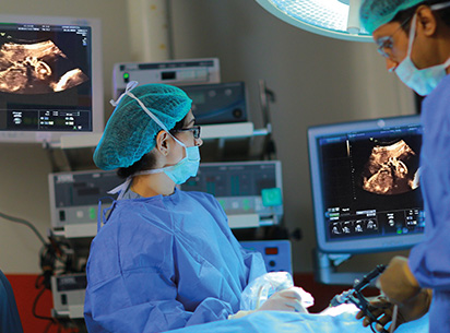
Advanced fetal treatment at CIMAR
It includes the entities of fetal therapy using means of laser, fetoscopy, minimally invasive fetal surgeries, EXIT procedures.
Fetoscopic laser
Fetoscopic laser or radiofrequency ablation using minimally invasive means is indicated especially in monochorionic pregnancy (shared placental blood vessels) complicated by TRAP, TTTS, Single fetal demise, TAPS.
Other conditions where minimally invasive techniques are done, which includes congenital cystic adenoid malformation, pulmonary sequestration, bladder outlet obstruction (inserting vesicoamniotic shunt).
EXIT (ex utero intrapartum treatment) is a specialized delivery procedure to secure the airway required to deliver babies who have airway obstruction eg. Neck tumors, bronchopulmonary sequestration, congenital cystic adenoid malformation.
All the procedures are highly skilled and require advanced training and expertise.